WHY HAVE A GLAUCOMA SCREENING?

|
Glaucoma is a progressive eye disease that can eventually cause permanent irreversible vision loss if left untreated. It doesn’t always cause noticeable symptoms in its early stages, so testing can help detect it before it progresses to a more advanced stage. It typically becomes more severe over time, which is why early diagnosis and management can significantly slow or even stop its progression. WHO IS AT RISK FOR GLAUCOMA? While anyone may develop glaucoma, some people are at a higher risk. Like with most medical conditions, understanding and identifying the risk factors, as well as timely treatment are key factors towards preserving health. Family history is an important factor for open-angle glaucoma, the most common type. Risk increases for those over the age of 50. Other risk factors include certain ethnic groups, a high degree of shortsightedness or farsightedness, previous eye trauma or injury, increased eye pressure, retinal detachment, health conditions such as diabetes and heart disease, and long-term use of certain steroid medications. WHAT ARE THE FIRST SIGNS OF GLAUCOMA?
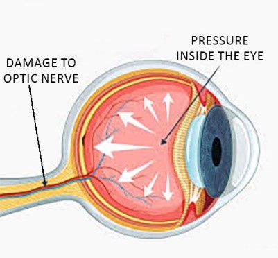
Glaucoma typically develops slowly with no early signs or symptoms, and most people do not notice vision changes until the condition has progressed and there is significant damage to the optic nerve. This is the reason it is usually first detected during an eye examination. There are several types of glaucoma, and not all of them are easy to identify in their earlier stages. The most common type of glaucoma is open-angle glaucoma which is due to inadequate drainage of fluid from the eye, resulting in increased intra-ocular pressure. It seldom causes noticeable symptoms in the early stages. As the disease progresses, a person may experience blurred vision, blind spots, difficulty seeing in conditions of low light and reduced peripheral vision. |
||
Less common is closed-angle glaucoma which tends to have more obvious early symptoms. Acute closed-angle glaucoma, as the name suggests, usually develops suddenly when drainage from the eye is blocked, leading to a rapid buildup of pressure against the optic nerve. Common symptoms include eye pain, headaches, eye redness, blurred vision and nausea. An acute attack is a medical emergency which requires immediate treatment. Early-onset glaucoma usually occurs before the age of 40, and hereditary factors tend to play a crucial role. Primary congenital glaucoma, which develops before the age of 3, is a rare childhood form of this eye condition with a genetic component. Normal-tension glaucoma is a unique type of glaucoma characterised by damage to the optic nerve despite normal eye pressure. While some may experience symptoms such as migraines, most people do not notice any symptoms in the early stages. |
||
|
WHAT TESTS ARE DONE TO DIAGNOSE GLAUCOMA? Glaucoma is a complex condition, and because no single eye test can provide enough information to make a diagnosis, a combination of tests is usually conducted. Special eye drops are used to dilate the pupils, allowing a clear view of the back of the eye and the optic nerve. For a few hours after pupil dilation, you may experience temporary blurry vision and light sensitivity. TONOMETRY – One of the main risk factors for glaucoma is raised pressure within the eye (intra-ocular pressure), but on its own this is not a definite diagnosis of glaucoma. It is possible to have glaucoma with lower eye pressure and not to have it with higher pressures. To test fluid pressure within the eye, a small amount of pressure is placed on the eye, either with a puff of air or tiny device. GONIOSCOPY – Fluid is constantly being produced in the eye and it flows out of the eye via the drainage angle. Gonioscopy examines the drainage angle and can determine if high intra-ocular pressure is caused by a closed, blocked or inadequately functioning angle.
OPHTHALMOSCOPY – The build-up of pressure eventually leads to damage of the optic nerve which conveys messages from the eyes to the brain. Ophthalmoscopy is the visual examination of the retina and optic nerve during a dilated eye examination when the nerve can be clearly seen. PERIMETRY - Glaucoma initially causes peripheral vision loss although the person may not be aware of this in the early stages of the condition. A perimetry test measures field of vision and is used to monitor changes to peripheral vision over time. PACHYMETRY – This test measures the thickness of the cornea, the clear membrane in front of the eye. A very thin cornea could indicate glaucoma or increase the risk of developing it. OPTICAL COHERENCE TOMOGRAPHY (OCT) - An OCT scan is an important test in diagnosing and monitoring glaucoma. It is used to measure the retinal nerve fibre layers around the optic nerve which is an important marker of early to moderate glaucoma damage. The OCT measurements help monitor any changes and progression of glaucoma over time and help to determine the management plan. WHAT IS THE OUTLOOK FOR PEOPLE WITH GLAUCOMA? Although there is no cure for glaucoma and any vision loss can’t be reversed, the condition is treatable. The aim of treatment is to reduce intra-ocular pressure and prevent further vision loss. With early detection, treatment and monitoring of glaucoma with regular eye examinations can slow or even stop its progression and help preserve vision. Visit your optometrist for a glaucoma screening. WORLD GLAUCOMA WEEK 8TH TO 14TH MARCH. |